An official website of the United States government




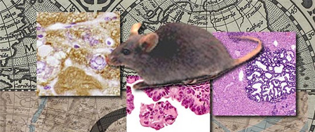
This Digitized Atlas of Mouse Liver Lesions provides color photomicrographs illustrating examples of some spontaneous, aging as well as chemically induced non-neoplastic and neoplastic histopathological hepatobiliary lesions commonly observed in B6C3F1 and Genetically Modified mice from acute, subchronic and chronic toxicity and/or carcinogenicity studies conducted by the National Toxicology Program . In addition to the color photomicrographs, content includes internationally accepted (standardized) diagnostic nomenclature as well of descriptions for some of the lesions.
Search by Pathology:
- Normal
- Congenital Lesions of the Liver
- Degenerative Lesions of the Liver
- Necrotic, Inflammatory and Vascular Lesions of the Liver
- Hyperplastic and Neoplastic Lesions of the Liver
- Miscellaneous Lesions of the Liver
- Toxicologic Lesions of the Liver
- Lesions of the Gallbladder
- Unique Liver Lesions of Genetically Altered Mice
Filter by First Letter:
A
- Adenoma
- Alb-uPA mice - hepatocellular adenoma
- Alb-uPA mice - nodular regeneration
- Alb-uPA transgenic mice
- AL-TAg mice
- AL-ras x AL-myc bitransgenic mice
- AL-TAgxAL-myc dual transgenic mice
- AL-TAgxAL-myc mice - anaplasia
- AL-TAgxAL-myc mice - biliary cysts
- AL-TAgxAL-myc mice - basophilic focus
- AL-TAgxAL-myc mice - bile duct adenocarcinoma
- AL-TAgxAL-myc mice - bile duct adenoma
- AL-TAgxAL-myc mice - bile duct proliferation
- AL-TAgxAL-myc mice - bromodeoxyuridine
- AL-TAgxAL-myc mice - cholangiocarcinoma
- AL-TAgxAL-myc mice - ductular formation
- AL-TAgxAL-myc mice - foci of cellular alteration
- AL-TAgxAL-myc mice - hepatocellular adenoma
- AL-TAgxAL-myc mice - hepatocellular carcinoma
- AL-TAgxAL-myc mice - hepatocellular neoplasm
- AL-TAgxAL-myc mice - hepatocytomegaly
- AL-TAgxAL-myc mice - karyomegaly
- Amyloidosis
- Anaplasia - AL-TAgxAL-myc mice
- Anaplasia of hepatocytes & oval cell proliferation
- Angiectasis
- Apoptosis
- Apoptosis - TGF alpha mice
- Apoptotic bodies
B
C
- Carcinoma in adenoma
- Chlordane
- Cholangiocarcinoma
- Cholangiocarcinoma - AL-TAgxAL-myc mice
- Cholangiocarcinoma - scirrhous reaction
- Cholangioma
- Cholecystitis
- Clear cell focus
- Coagulation necrosis
- Congenital lesions of the liver
- Crystalline inclusions - gallbladder
- Cyst - gallbladder
- Cystic degeneration
- Cytomegaly
- Cytomegaly associated with chlordane exposure
- Cytomegaly associated with Helicobacter
- Cytomegaly of hepatocytes & oval cell proliferation<</a>
- Cytoplasmic alteration
- Cytoplasmic vacuolation
D
E
F
G
- Gallbladder
- Gallbladder - crystalline inclusions
- Gallbladder - cyst
- Gallbladder - ectopic pancreas
- Gallbladder - hyaline degeneration
- Gallbladder - hyperplasia
- Gallbladder - inclusions
- Gallbladder - inflammation
- Gallbladder - papillary adenoma
- Gallbladder - papillary hyperplasia
- Genetically altered mice
- Giant cells in histiocytic sarcoma
- Glycogen Deposition
H
- HBV transgenic mice
- HBV transgenic mice - hepatic neoplasia, gross
- HBV transgenic mice - hepatocellular adenoma
- Helicobacter hepaticus
- Hemangioma
- Hemangiosarcoma
- Hematopoiesis
- Hepatic atrophy with Ito cell tumors
- Hepatitis
- Hepatitis B
- Hepatoblastoma
- Hepatocellular adenoma
- Hepatocellular adenoma - Alb-uPA mice
- Hepatocellular adenoma - AL-TAgxAL-myc mice
- Hepatocellular adenoma - HBV transgenic mice
- Hepatocellular adenoma - TGFalpha mice
- Hepatocellular carcinoma
- Hepatocellular carcinoma - AL-TAgxAL-myc mice
- Hepatocellular hypertrophy
- Hepatocholangiocarcinoma
- Hepatocyte atrophy with hemangiosarcoma
- Hepatocyte degeneration in fatty change
- Hepatocytomegaly - AL-TAgxAL-myc mice
- Hepatocytomegaly - TGF alpha mice
- Hepatodiaphragmatic nodule
- Hepatopathy
- Histiocytic sarcoma
- Histiocytosis
- Hyaline bodies
- Hyaline change
- Hyaline degeneration
- Hyaline degeneration - gallbladder
- Hyperplasia - gallbladder
- Hyperplastic lesions of the liver
I
K
L
M
- Malignant lymphoma
- Malignant melanoma
- Mast cell tumor
- Melanoma
- Metastatic tumor, ovarian teratoma
- Metastatic tumor, malignant melanoma
- Metastatic tumor, lymphoma
- Microgranuloma
- Microinvasion
- Microvesicular fatty change
- Miscellaneous lesions of the liver
- Mixed focus
- Myelodysplasia - Tg.AC mice
- Myelopoiesis
- Myeloproliferation - Tg.AC mice
N
O
P
- Papillary adenoma - gallbladder
- Papillary hyperplasia - gallbladder
- Peliosis associated with hemangiosarcoma
- Peliosis hepatis
- Perisinusoidal lining cells
- Periodic acid Schiff stain
- Pigment - toxic change
- Ploidy
- Polyploidy
- Polyploidy associated with apoptosis
- Polyploidy associated with hepatocyte hypertrophy
- Pseudoinclusion with oval cell hyperplasia
S
T
- Telangiectasis
- Tension lipidosis
- Teratoma
- Tg.AC mice
- TGF alpha mice - apoptosis
- Tg.AC mice - myeloproliferation
- Tg.AC mice - myelodysplasia
- Tg.AC mice - erythroleukemia
- TGF alpha
- TGF alpha mice
- TGF alpha mice - hepatocellular adenoma
- TGF alpha mice - hepatomegaly
- TGF alpha mice - karyomegaly
- Torsion
- Toxic change
- Toxic change - Kupffer cell hyperplasia
- Toxic change - oval cell hyperplasia
- Toxic change - pigment
- Toxicologic lesions of the liver
- Trabecular carcinoma
- Transgenic mice
- Transgenic mice - Alb-uPA mice
- Transgenic mice - AL-TAg mice
- Transgenic mice - AL-TAgxAL-myc
- Transgenic mice - AL-ras x AL-myc
- Transgenic mice - HBV
- Transgenic mice - hepatocellular adenoma
- Transgenic mice - Tg.AC mice
- Transgenic mice - TGF alpha
U
V
Contact