 An official website of the United States government




The Digitized Atlas of Mouse Liver Lesions

Much of the work carried out by DTT is in support of the National Toxicology Program (NTP), an interagency partnership of the Food and Drug Administration, National Institute for Occupational Safety and Health, and NIEHS.
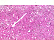
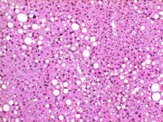
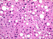
Fatty change, mild. The circular clear spaces represent areas previously occupied by fat in this mouse on a choline deficient diet. The fat is dissolved out by xylene during processing of the tissue.
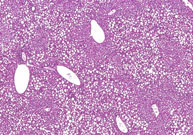
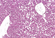
Periportal fatty change. The large, sharply delineated, clear vacuoles represent fat that has been dissolved during tissue processing. Deposition of fat in the hepatocyte cytoplasm often displaces the hepatocyte nucleus to the periphery of the cell.
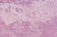
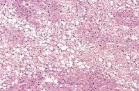
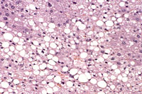
Large areas of fatty change without a distinct lobular pattern are present in this liver. The large, sharply delineated, clear vacuoles represent macrovesicular fat that has been dissolved during tissue processing. Deposition of fat in the hepatocyte cytoplasm often displaces the hepatocyte nucleus to the periphery of the cell.
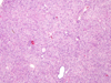
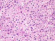
This represents an example of microvesicular fatty change. Multiple small vacuoles present within the hepatocytes give the cytoplasm a foamy appearance.
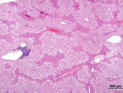
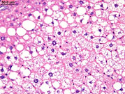
This example of fatty change represents a combination of microvesicular and macrovesicular fatty change with a distinct lobular pattern. The macrovesicular fat is localized at the periphery and probably represents condensation of microvesicular vacuoles.
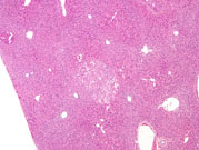
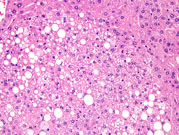
This is an example of focal fatty change with affected hepatocytes containing a mixture of macrovesicular and microvesicular fat.
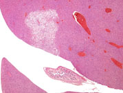
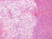
A specific example of focal fatty change seen in mice and present in the median lobe represents tension lipidosis when it is near the falciform ligament and gallbladder.
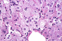
Fatty change (small lipid droplets) in the liver of a mouse given ethionine.
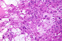
Cytoplasmic vacuolation of hepatocytes (fat) in a mouse given benzene hexachloride. Vacuoles vary from small globules to large vacuolated areas in the cytoplasm.