 An official website of the United States government





Much of the work carried out by DTT is in support of the National Toxicology Program (NTP), an interagency partnership of the Food and Drug Administration, National Institute for Occupational Safety and Health, and NIEHS.
These examples represent portions of the liver that protrude through the diaphragm.
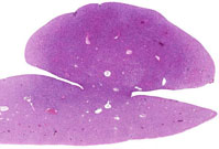
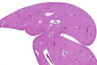
Low magnification examples of hepatodiaphragmatic nodules in rats. These represent portions of the liver that extend into the thoracic cavity and are covered by a thin, fibrous portion of the diaphragm. These are rare in mice and less nodular compared to rats.
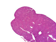
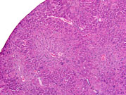
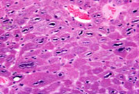
Low, medium and high magnification of a hepatodiaphragmatic nodule in a mouse. Note the characteristic elongated pattern of the nuclear chromatin in the nodule.